LONG CASE - PRACTICAL
This is an online e log book to discuss our patient de-identified health data shared after taking his / her / guardians signed informed consent. Here we discuss our individual patients problems through series of inputs from available global online community of experts with an aim to solve those patients clinical problem with collective current best evident based input.
This E blog also reflects my patient centered online learning portfolio and your valuable inputs on the comment box is welcome.
A 51 year old male presented with. Pleural effusion with Liver abscess.
I have been given this case to solve in an attempt to understand the topic of " patient clinical data analysis" to develop my competency in reading and comprehending clinical data including history, clinical findings, investigations and come up with diagnosis and treatment plan.
CASE:
A 51 year old patient who is a resident of chitayala, who works as a labourer in a goods company came to the hospital with chief complaints of:
• Fever since 10 days
• Shortness of breath since 10days
• Cough since 7 days
HISTORY OF PRESENT ILLNESS :
The patient was apparently assymptomatic 10 days back. Then he developed high grade fever which was insidious in onset associated with chills and rigours and was relieved on taking medications. It was associated with cough and shortness of breath.
The patient was able to walk a distance of 1km 10 days back and slowly started developing shortness of breath on walking for short distances, which became more severe that he has SOB even at rest.
no Orthopnea
no paraxsomal nocturnal dyspnea
no pedal Edema.
Cough since 7 days which is productive, mucoid in consistency, whitish, scanty in amount, non foul smelling, non blood stained, aggrevated during night time and on supine position.
Right sided chest pain - diffuse, intermittent, dragging type, aggravated on cough, non radiating, not associated with sweating and palpitations .
Weight loss - present
no loss of appetite
no history of pain abdomen
No abdominal distension, vomiting, loose stools.
no history of burning micturition .
PAST HISOTRY:
History of jaundice 20 days back which resolved in a week without any medications.
No H/O DM/HTN/TB/CVA/CAD/COPD/epilepsy
FAMILY HISTORY:
No similar complaints in the family
PERSONAL HISTORY:
patient is a chronic smoker, smokes a pack of cigarettes since past 25 years.
He is a chronic alcoholic consumes 325ml (quarter ml of whiskey) daily.
Sleep - adequate
no bowel and bladder disturbances.
PROVISIONAL DIAGNOSIS:
51 year old with fever, cough and SOB with provisional diagnosis as:
1-pleural effusion
2-pneumonia
3-tuberculosis
GENERAL EXAMINATION :
Patient is conscious, coherent and cooperative, moderately built and nourished .
No signs of pallor, cyanosis, clubbing, icterus, koilonychia, lymphadenopathy, pedal edema .
VITALS:
Temp : afebrile
PR : 83bpm ,normal volume, regular rhythm, normal character, no radio-radial delay.
BP : 110/70 mmHg, measured in supine position in both arms .
RR - 22cpm
SYSTEMIC EXAMINATION :
Patient examined in sitting position after taking consent in a well lit room.
ORAL CAVITY:
Nicotine staining seen on teeth and gums.
RESPIRATORY SYSTEM:
INSPECTION:
•Respiratory movements appear to be decreased on right Side
•Shape of chest: barrel
•Trachea is shifting towards left
•Nipples are in 4th Intercoastal space
•Apical impulse visible in 5th intercostal space.
•no dilated veins, scars, sinuses, visible pulsations
•no rib crowding,
•no accessory muscle usage .


PALPATION:
•No local rise of temperature
•No tenderness
•All inspiratory findings are confirmed
•Trachea is shifted to left
•Apical impulse - in left 5th ICS, 1cm medial to mid clavicular line
•Respiratory movements decreased on right side
•Tactile and vocal Phremitus - reduced on right side in mammary, infra-axillary and infrascapular region.
•AP diameter : 32cm
•Transverse diameter : 26cm
•AP:T ratio - 1:2
•Chest circumference : 9.5 cm expiratory
9.8 cm inspiratory
PERCUSSION : Right Left
Supraclavicular. Resonant. Resonant
Infraclavicular. Resonant. Resonant.
Axillary. Dull. Resonant
Infra-axillary. Dull. Resonant
5th ICS. Dull. Resonant
Suprascapular. Resonant. Resonant
Interscapular. Dull. Resonant
Intrascapular. Dull. Resonant
AUSCULTATION: Right. Left.
Supraclavicular. NVBS. NVBS
Infraclavicular. NVBS. NVBS
Mammary. ⬇️. NVBS
Axillary. NVBS. NVBS
Infra-axillary. ⬇️. NVBS
Suprascapular. NVBS. NVBS
Interscapular. ⬇️. NVBS
Intrascapular. ⬇️. NVBS

OTHER SYSTEMS:
GASTROINTESTINAL SYSTEM :
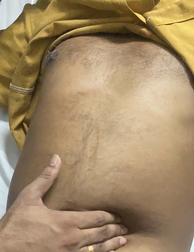
INSPECTION:
Abdomen - distended
All quadrants of abdomen are equally moving with respiration except Right upper quadrant. No visible, No visibe sinuses ,scars, visible pulsations or visible peristalsis
PALPATION:
All inspectory findings are conformed
No tenderness .
Liver - is palpable 4 cm below the costal margin and moving with respiration.
Spleen : not palpable.
Kidneys - bimanually palpable.
PERCUSSION - normal
AUSCULTATION - bowel sounds heard, No bruits.
CVS :
S1 and S2 heard. no murmurs
CNS : NAD
Video:
INVESTIGATIONS :
XRAY:
ELLIS curve (s shaped curve/Damoiseaus curve): curved shadow at the king base, blunting the costophrenic angle and ascending towards the axilla. Shifting dullness is seen on examination.

ECG:


Colour - straw coloured
Total count -2250 cells
DLC - 60% Lymphocyte, 40% Neutrophils
No malignant cells.
Pleural fluid sugar = 128 mg/dl
Pleural fluid protein / serum protein= 5.1/7 = 0.7
Pleural fluid LDH / serum LDH = 0.6
INTERPRETATION:
Exudative pleural effusion.
Serology - negative
Serum creatinine - 0.8 mg/dl
CUE - normal
CT SCAN:


FINAL DIAGNOSIS :
1. Right sided pleural effusion
2. Right lobe liver abscess
TREATMENT :
Inj. PIPTAZ 2.5gm iv QID
Tab. AZITHROMYCIN 500 OD
Inj. METROGYL 100ml TID
Tab. DOLO 650mg
Inj. NEOMOL 1gm IV
O2 inhalation
IV fluids: normal saline
Inj optineuron
Temperature chart 4 hrly
Bp, Sp02 chart 4hrly
Inj.AMIKACIN iv BD


Comments
Post a Comment